
 Se Habla Español
Advanced Dental Technology & Services Lancaster
Using the Latest Tools for High-Quality Dentistry
Dentistry is a constantly growing and evolving field, and as such, it takes dedication to learning about the latest technology to stay on the cutting edge of high-quality care. Our doctors have made it a point to make sure that Lancaster Family Smiles uses state-of-the-art instruments and techniques for accurate diagnoses and precise procedures. The next time you call our dental office in Lancaster for an appointment, feel free to ask about some of the advanced tools we use to protect and treat your smile.
Committed to Comfortable, Productive Dental Experiences
- Highly Accurate CT Scans for Precise Implant Placement
- Shorter Healing Time with L-PRF Machine
- Faster, Safer Digital X-Ray Technology
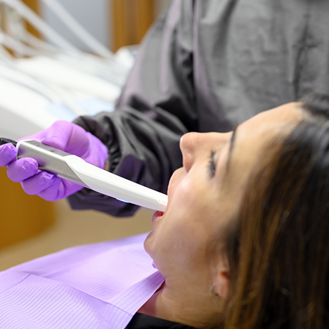
Intraoral Camera
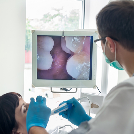
When we want you to see what’s going on inside your mouth, we’ll use a wand-like device called an intraoral camera. It can capture images inside your mouth without external lighting, but its most useful feature is that it can be connected to a computer so that we can show you exactly what we’re seeing. This way, you’ll have a clear idea of exactly what the problem is and why we’re recommending a specific form of treatment.
Digital X-Rays
Dental problems aren’t always visible in your teeth and gums – they can also be lingering under the surface of your smile, growing worse without you even realizing what’s happening. Here in Lancaster, our team is dedicated to diagnosing these conditions as soon as possible for the sake of your comfort and safety. Thankfully, our digital X-ray technology allows us to capture exceptionally detailed images of your oral structures and use them as valuable tools for the treatment and recovery process.
Many older patients are familiar with traditional X-rays, as well as their many pitfalls. These photographs had to be developed slowly in a darkroom with hazardous chemicals and then stored in bulky filing cabinets. The amount of radiation a patient had to be exposed to was also no laughing matter. Our digital system transforms this process for the better, reducing the necessary radiation by up to 90% and shortening the development process to mere seconds. The images are pulled up easily on our computer system and color-coded to help our patients understand what they’re looking at!
CT/Cone Beam Scanner
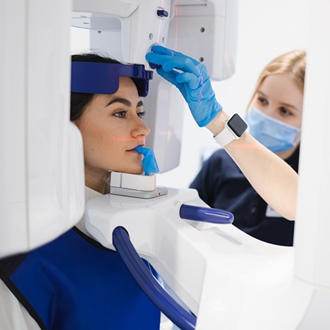
As useful as regular dental X-rays are, they’re not always enough. For certain procedures, we need to use a dental cone beam CT scan. The machine rotates around your head and takes multiple pictures that form a 3D image. This gives us a complete view of your teeth as well as the various soft tissues, nerve paths, and bones in the craniofacial region. All of this information makes it easier for us to plan specific procedures.
L-PRF Machine
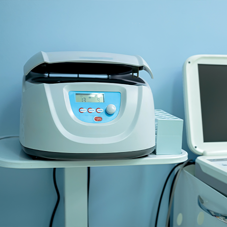
It’s important for your mouth to heal properly after a procedure. To help the process along, we can apply platelet-rich fibrin, or PRF, to sites where dental surgery has been performed, encouraging the hard and soft tissues to repair themselves sooner. PRF is derived from your own blood using a state-of-the-art L-PRF machine, which draws out the fibrin from blood plasma using centrifugal force. You can ask our doctors for more details about how PRF is used in dentistry.
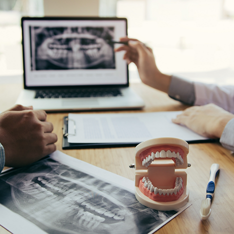
Digital Impressions
Accurate and comfortable restorations are essential if you want your smile to appear uniform and remain healthy. This is why our dentists use a digital impression system. By scanning your teeth and gums, the device produces a three-dimensional model that appears on a chairside monitor so you and our team can easily view the image. After making the necessary adjustments, we send the digital model to lab technicians to start the fabrication process.
About Us Meet the Dentists Tour Our Office View Our Services